Revamping the Digital Interface
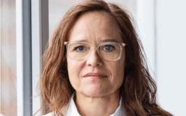
Sitting Down With… David Wilbur, Former Director of Cytopathology and Clinical Imaging at Massachusetts General Hospital and current Chief Medical Scientist at Corista LLC, USA

Why has the US been slower to adopt digital pathology?
Although there is no question about the versatility and accuracy of digital pathology, there is a question surrounding its business case; most departments consider it a cost rather than a revenue-generating concept. European institutions have an advantage over those in the US because their health care systems incorporate digital pathology into their plans and fund these projects over a period of time. Here, we have to demonstrate that it can generate revenue, provide efficiency to save money, or improve patient safety before allocating the money and resources.
I believe that one of the main things holding back the adoption of digital pathology is the poor user interface between the pathologist and the viewing station – something that I have been trying to address over the last 15 years. As the product of over a century of development and fine-tuning, the microscope is exceedingly efficient and ergonomic for the user, and this is difficult to replicate in digital viewing systems. Inspired by the “Powerwall” at Leeds, which is a fantastic way to view digital slide images, I wanted to translate the way pathologists handle slides under the microscope into digital systems.
That’s where the laser box virtual slide stage comes in. An artificial slide on top of a small, laser-controlled platform allows you to move the digital image around on the screen as if it were a real glass slide. We have successfully tested this prototype and pathologists have consistently commented on the improved efficiency of the technique compared with using a mouse, which may be excellent for navigating a computer screen, but is quite slow and laborious for diagnostic review of a whole slide image. At the moment the laser box only works with the Corista viewing system but, in the future, I would like to see it as a piece of standalone equipment that can be plugged into any system.
What do you hope to achieve as Chief Scientific Officer at Corista?
In addition to optimizing the interface between pathologists and digital viewing systems, my ambition is to further the potential applications of artificial intelligence (AI). Our first foray into AI has been in screening renal biopsies to identify glomeruli. Although that sounds like an arcane task, for a renal pathologist who has to find and evaluate every glomerulus on a biopsy, the ability to navigate to those glomeruli immediately is highly desirable. In addition, our work includes the registration of all special stains, meaning that the same glomerulus is co-located on each special stain for digital review simultaneously. We’re now striving to go one step further and classify the glomeruli based on diagnostic annotations that renal pathologists have given us. If successful, we will then be able to apply those same tools to a variety of biopsies such as prostate, breast, GI, and urinary tract. Through digital “prescreening,” we hope to address the sorts of scenarios that will make the pathologist much more efficient and potentially more accurate.
What’s your outlook on the future of pathology?
There are a lot of naysayers who believe that pathology is going to disappear because of AI, but I am of the firm belief that it will make us far more productive. Genitourinary (GU) pathologists, for example, will use AI to pre-screen prostate biopsies on arrival to identify “hotspots,” or areas of high probability that the pathologist needs to examine most closely. Many of these areas will be false positives, but that isn’t a problem – the key part is that there will be a pathologist to look at those slides and discriminate between cancer and benign mimics. This guided screening will mean that, instead of only having time to look at a handful of prostate biopsies in a morning, GU pathologists will be able to look at many more, greatly improving efficiency.
What is the proudest moment of your career?
Interestingly enough, that moment actually happened a few months ago when my son – a practicing radiologist – and I led a workshop together at the American Society of Cytopathology’s annual meeting. It was a pathology and radiology correlation conference that we ran as a seminar; my son presented the radiology part on one side of the room and cytologists presented the pathology from the other side. We talked about differential diagnoses from both the pathology and radiology aspects, showing how important it was for patient care to have both perspectives. Seeing my son take his place as a teacher and clinician really was an incredible experience.
While completing my undergraduate degree in Biology, I soon discovered that my passion and strength was for writing about science rather than working in the lab. My master’s degree in Science Communication allowed me to develop my science writing skills and I was lucky enough to come to Texere Publishing straight from University. Here I am given the opportunity to write about cutting edge research and engage with leading scientists, while also being part of a fantastic team!
