OCT for the People
Can a low-cost OCT device aid biomedical research and improve disease diagnosis in rural and low-income settings?
Adam Wax |
At a Glance
- OCT is an effective tool for biomedical research and for the diagnosis of a range of retinal diseases – but it’s far from inexpensive
- A 3D printed approach can produce a portable OCT device for a fraction of the cost, helping to expand the reach of OCT
- There are drawbacks, such as the number of images you can take per second – but the device is still an effective diagnostic and imaging tool
- Working to democratize previously expensive medical equipment, such as OCT, ultimately brings better healthcare to all.
I’ve been working in the field of biomedical optics for 20 years and, in that time, I’ve brought a number of different products through clinical trials and to market – mostly endoscopes. In general, I’ve been frustrated with how expensive it is to bring a new medical product to market. By the time you’ve completed research and clinical trials, you end up with a device that could cost upwards of a hundred thousand dollars, and costing up to a thousand dollars per use. And that means that it’s very tough to get new technologies adopted. In short, we do a great deal of work, spend a lot of money, but end up with very few products that actually bring enough benefit to convince the market to buy them.
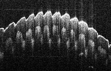
An image of a fingerprint taken using the OQ LabScope
Coming from a physics background, I’ve always loved how optical approaches can provide very elegant and precise descriptions of scientific phenomena. Taking this technology and applying it to biology and medicine, a much messier field, is where the biggest challenges lie. Nevertheless, I was inspired to start a company with a different vision: to prove that we can create low-cost, high-quality biomedical imaging products, and still make a profit. Our first project? Optical coherence tomography (OCT).
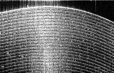
An OCT image of scotch tape, showing over 40 layers of tape and adhesive.
An elegant – but expensive – approach
OCT is already an established technology with known benefits – it provides noninvasive, cross sectional imaging, and penetrates more deeply into tissue than confocal microscopy, without requiring a fluorescent reporter. Essentially, it’s an optical analog of ultrasound. In ultrasound, you send a pulse of sound into your sample, and wait for it to ‘bounce’ back. The time it takes tells you how deep it’s gone. With OCT you use light, which moves much more quickly – so you can’t just time it with an electronic stopwatch! Instead we do interferometry: we break some of the light off into a reference arm, send it down a reference path, and when that light comes back it’s matched with the light that has gone into the sample, giving you an interference pattern. By varying the length of the reference arm, you can map out a depth-resolved reflection profile from your sample. And you can get beautiful detail on all the cross sectional layers.
These benefits have led OCT to become the current gold standard in detecting diseases of the retina – it’s a great approach for detecting diseases like diabetic retinopathy, age-related macular degeneration (AMD), and glaucoma. The main problem is the expense of OCT systems, which means that they are more typically found in big eye centers; if you’re 100 miles away from the nearest city, you don’t have easy access to the technology. And if you’re in a low- or middle-income country, the problem is the same.
We set out to create a lower cost device to increase access to this imaging technology. The result is a device about the size of a shoebox that costs under $10,000, which essentially allows doctors to visit remote locations or perhaps a retirement home, where patients are less mobile, and scan 30 or 40 people in a single session.
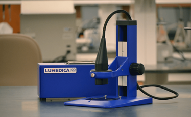
The OQ LabScope OCT device
Mice, materials and medicine
But before we can sell our device for human use in the US, we need FDA approval. Right now, we have a device on the market, OQ LabScope, for research and educational purposes. So far we’ve seen good uptake, especially by those who never thought about using OCT as it was simply too expensive. We’ve had biomedical researchers trying it on many different organ sites – for example, researchers interesting in imaging different parts of the mouse, from the eyes, to the skin or the teeth. We’ve had interest from pathologists who want to look at tissue sections on the lab bench, to figure out where to take their histological sections. We’ve also seen interest from people working on materials research. Say you’re working on a new coating for a phone or television screen, or on optical fibers – our device offers a nondestructive testing option. There aren’t a lot of imaging options for this type of work, so our relatively cheap and portable device, which can be easily shared among researchers, is getting a positive response.
Lowering the cost of the device dramatically was our main challenge – and the beautiful thing about the device, for us, is the design of the spectrometer which is the heart of the OQ LabScope. Usually, spectrometers are built from high tolerance parts that are locked down so they never move. But we took a different approach and decided to let our spectrometer “breathe” while still staying aligned; we built it with 3D printed parts that are pushed into place and glued by hand. When we ship it across country and it’s taken out of the box it’s already aligned – there’s no need for a highly skilled expert to come in and calibrate it for you. It’s “plug and play.” Additionally, for many of these devices you need to have a separate computer and screen. Our device doesn’t need that – the computer is inside the system so you can plug it into a monitor, or you can use a tablet or cell phone to control it, keeping the footprint really small and the costs down.
However, there are trade-offs when developing a cheaper alternative. We meet nearly all of the specifications for standard OCT systems in terms of resolution and signal-to-noise ratio. However, our device is a bit slower – we’re limited to about 9,000 A-scans per second which is about 20 images per second, while more sophisticated machines can take perhaps 20,000–30,000, or even as many as 50,000 A-scans per second, or 40-60 frames per second. If you’re trying to image a really fast process, our device may not be the best option, but for many applications this slower image rate isn’t a problem. The aim isn’t to produce the most sophisticated OCT machine available, it’s to aid research, increase access – and, eventually, to help catch disease early.
In fact, the push for incremental advances in current OCT technology is one of the reasons why it remains so expensive – if you’re pushing to get another three or five percent performance, you can end up with a much more expensive instrument, with features that don’t necessarily aid basic functions like diagnosis. Our aim is to strip all that back and democratize OCT.
A vision for the future
There was a time that ultrasound was only found in big hospitals, but now there are portable ultrasounds that they take out on ambulances when there’s an emergency call. We hope that our version of OCT will have that kind of portability, and provide that level of access.
Our next goal is to get a retinal imaging product on the market. We’re working toward FDA clearance and a CE mark. We’re also looking at the international market, especially in areas where access to OCT is more limited, such as India and China. Ultimately, we believe that a low-cost instrument could help save the sight of millions of people. And it doesn’t need to stop at OCT – cheaper, portable alternatives to expensive medical equipment could make a huge difference in all areas of healthcare, bringing the latest diagnostic tools to many more patients.
Adam Wax is a Professor of Biomedical Engineering at Duke University, North Carolina, USA, and President and Chief Scientist of Lumedica, Inc.
For more information about the LabScope, visit Lumedica.co or email [email protected].
Adam Wax is a Professor of Biomedical Engineering at Duke University, North Carolina, USA, and President and Chief Scientist of Lumedica, Inc.


