What?
Tracking tiny tumors in real time could soon be a reality, thanks to a new nanotechnology-based approach for following the spread of micrometastases (1).
Who?
A team of researchers from Rutgers University and Cancer Institute of New Jersey, USA, and Singapore University of Technology and Design.

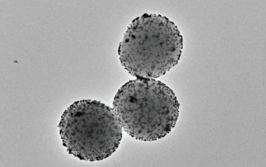
Figure 1. In the study, human breast cancer cells in a mouse model were “chased” with novel rare earth nanoscale probes injected intravenously. When the subject is illuminated, the probes glow in an infrared range of light that is more sensitive than other optical forms of illumination. In this image, the probes show the spread of cancer cells to adrenal glands and femur bones.
Image credit: Harini Kantamneni and Professor Prabhas Moghe, Rutgers University New Brunswick
Why?
Current imaging methods cannot spot tumors in the early stages of metastases, confounding cancer staging and treatment decisions – and the “Achilles heel” of surgical management, say the authors.
How?
Light-emitting nanoparticles are injected intravenously and reveal lesions that can’t be detected using contrast-enhanced MRI, allowing real-time surveillance of cancer spread in a mouse model (see Figure 1).
When?
According to the authors, the technology could be available in the clinic within five years, and lead to more accurate cancer monitoring.
- H Kantamneni et al., “Surveillance nanotechnology for multi-organ cancer metastases”, Nat Biomed Eng, 1, 993–1003 (2017).

I have an extensive academic background in the life sciences, having studied forensic biology and human medical genetics in my time at Strathclyde and Glasgow Universities. My research, data presentation and bioinformatics skills plus my ‘wet lab’ experience have been a superb grounding for my role as a deputy editor at Texere Publishing. The job allows me to utilize my hard-learned academic skills and experience in my current position within an exciting and contemporary publishing company.


