Imaging Innovation (The Art of Translation)
New tools and techniques are giving us a close-up view of cells and organs.
Multimodality Imaging
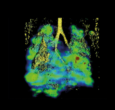
In a metastatic model of melanoma, a lung CAT scan (in solid yellow) is fused with a SPECT image highlighting metastatic lesions (blue-to-red to indicate lesion density).
Credit: National Cancer Institute
RNA at the Nanoscale
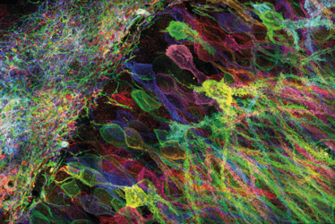
MIT researchers have developed a newway to image proteins and RNA insideneurons of intact brain tissue, using expansion microscopy.
Credit: Yosuke Bando, Fei Chen, Dawen Cai, Ed Boyden, and Young Gyu
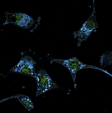
Smart Probes
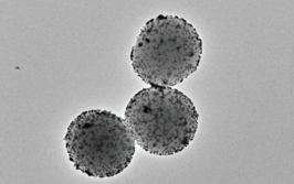
SmartFlare Detection Probes, developed at Northwestern University, isolate Nodal-positive melanoma cells from a heterogeneous population. The Nodal-positive cells (blue) are also positive for CD-133 (green), another biomarker associated with cancer stem cells and drug resistance.
Credit: National Cancer Institute
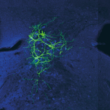
Mind Map
Scientists at Columbia University have developed a new tool to probe the activity and circuitry of neurons in the mouse brain, by tracking the spread of a modified version of rabies virus.
Credit: Andrew Murray
