Babies’ Breath
New research reveals a potential noninvasive predictor of bronchopulmonary dysplasia risk in preterm infants
At a Glance
- Preterm infants, especially those requiring prolonged oxygen therapy, are at risk of developing bronchopulmonary dysplasia
- None of the proposed genetic or cytokine biomarkers for BPD risk have been replicated upon closer study
- A new type of biomarker – mitochondrial function – may be more successful, and can be noninvasively tested in umbilical cord blood cells
- If validated, mitochondrial function testing could help doctors determine which infants need modified respiratory support
Many prematurely born infants struggle with breathing, and as many as half may develop bronchopulmonary dysplasia (BPD), a lung function abnormality that stresses the infants’ underdeveloped lungs and can result in lifelong chronic or even fatal disease. Reactive oxygen species (ROS) arising from prolonged oxygen therapy in babies who are unable to breathe sufficiently on their own interfere with the lungs’ maturation and may mean that the terminal saccules – vital for gas exchange during breathing – don’t develop correctly. But is there any way to predict which preterm infants may develop BPD, and therefore, which might need modified respiratory support? Until recently, the answer has been “no” – but a new type of biomarker may change that.
Why we did it
Over the last few years, many medical professionals have hypothesized that pulmonary vascular dysfunction may be an important causative factor in the development of BPD in preterm infants. Even though evidence has now emerged regarding the central role of mitochondria in hyperoxia-related tissue injury – a key pathogenic factor for prematurity-related pulmonary disease – mitochondrial function is a relatively novel and under-investigated area in this disease process.
Human umbilical venous endothelial cells (HUVEC) have often been used as model systems to study the role of vascular function and pathology in the pathogenesis of several diseases, including diabetes, atherosclerosis and hypertension. But, until our study, they had never been used to investigate endothelial function as a risk factor for diseases to which the infants from whom they are obtained are susceptible in their earliest days.
We have collaborated in the past with our co-investigators at the University of Alabama’s Department of Pathology on a project that used HUVEC obtained from term newborn infants to investigate mitochondrial bioenergetic differences arising from ethnicity. As a neonatal physician and a lung development and injury researcher, the approach intrigued me – and, as a result, I conceived the idea of using HUVEC obtained from preterm infants to measure endothelial function. My goal was to compare function between infants who later developed lung disease or died early versus those who survived without BPD. I was especially lucky to work at the University of Alabama at Birmingham, one of the few centers in the United States that has all the necessary components to work collaboratively on such a project – researchers with expertise in the areas of lung development and injury and in mitochondrial and redox biology research, as well as neonatal physicians and a robust and large neonatal intensive care unit. In the end, our study spanned four years, and I’m grateful to all of the people involved; without such a broad range of skills, we could never have completed our work.
Why could the results of the project be so revolutionary? At the moment, most scoring systems that predict BPD risk rely on variables like gestational age and birth weight differences, which contribute significantly to the developmental immaturity that places preterm infants at risk of complications. Those aren’t always reliable measures, though, so researchers have made numerous attempts to identify potential biomarkers of these infants’ risk of lung disease. Several studies have suggested various cytokines as possible biomarkers; others have proposed genetic polymorphisms (1)(2). Unfortunately, no such link has been replicated in subsequent studies. In short, there has been no single reliable biomarker for predicting an individual’s risk of developing lung disease. That’s why our new discovery – that bioenergetic function (measured in cells that are relatively easy to obtain from preterm infants at the time of their birth) may be a marker for their risk of BPD – is so important. And, if successfully validated, it could improve our ability to identify prematurely born infants at increased risk before they develop significant lung injury.
How we did it
In our study, we harvested HUVEC from the umbilical cords of 69 infants born at or earlier than 32 weeks’ gestational age. We carried out bioenergetics measurements (intact HUVEC oxygen consumption) with a flux analyzer, as well as reactive oxygen species (ROS) measurements in hyperoxia-exposed HUVEC using fluorescence-based methods. Finally, we used quantitative PCR to measure damage to mitochondrial DNA.
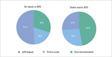
Ultimately, we identified HUVEC bioenergetic function (measured as basal and maximal oxygen consumption under standard assay conditions) as the most significant factor that could reliably distinguish between infants with and without BPD. Fortunately, there are already several platforms that can reliably measure cellular oxygen consumption using as few as 1,000 cells – and we’re currently in the process of developing a protocol that will use such systems to validate HUVEC bioenergetic measurements as a biomarker for BPD risk. The test won’t hit the clinic tomorrow – validation will likely take at least two to three years – but, if successful, it would help us avoid the need for cell culture and allow us to measure endothelial mitochondrial bioenergetic function in primary cells obtained directly from the umbilical cords of infants at the time of their birth.
It’s possible that, in the future, we might be able to test preterm infants for mitochondrial dysfunction in other ways. Mitochondrial genetic inheritance occurs through maternal transmission, so one particularly interesting approach would be test mitochondrial function and genetic differences using cells obtained from pregnant mothers, especially those at increased risk of preterm delivery. Sampling for this wouldn’t be difficult; buccal epithelial cells obtained through oral mucosal scrapings would suffice. It’s also possible to obtain human umbilical arterial endothelial cells (in addition to the HUVEC we used) from every newborn infant without the need for invasive procedures. These cells could also serve as a source of information regarding mitochondrial function and genetics.
Of course, along with the clinical test will come the question: who should be tested? In my opinion, any infant who is at risk for lung injury because of premature birth is likely to benefit from endothelial mitochondrial function testing. This is especially true for the various subgroups of infants that we particularly identified as having bioenergetic and redox dysfunction in our study – namely, those exposed to maternal and placental infection or inflammation and African-American infants.
What’s next?
Our study identifies an association between BPD risk in preterm infants and the degree of their endothelial mitochondrial dysfunction. However, the mechanisms behind that association are still unclear and need further investigation. We have some preliminary hypotheses – for instance, endothelial mitochondrial dysfunction could cause deranged pulmonary angiogenesis by reducing nitric oxide and vascular endothelial growth factor availability. Then, the increased ROS generation from these cells could lead to the dysfunction of neighboring cells that constitute the pulmonary tree.
Because mitochondrial bioenergetic function depends on proteins in the electron transfer chain, which are derived from mitochondrial and nuclear gene expression, it’s important to investigate both of these sets of genes. Variations in mitochondrial genetic haplotypes, differences in interactions between these two genomes (“mito-Mendelian genetics”), or both could impair or modify mitochondrial response to hyperoxia. Additionally, we also need to investigate mitochondrially targeted therapeutic strategies that could decrease pulmonary mitochondrial dysfunction – and thereby potentially also reduce the risk of lung injury in preterm infants. With a combination of better tests and better treatments, these babies may soon be able to breathe more easily.
Jegen Kandasamy is Assistant Professor at the University of Alabama at Birmingham and Director of the Rare Disease and Congenital Anomalies Programs at Children’s Hospital of Alabama, Birmingham, USA.
- CV Lal, N Ambalavanan, “Biomarkers, early diagnosis, and clinical predictors of bronchopulmonary dysplasia”, Clin Perinatol, 42, 739–754 (2015). PMID: 26593076.
- CV Lal, N Ambalavanan, “Genetic predisposition to bronchopulmonary dysplasia”, Semin Perinatol, 39, 584–591 (2015). PMID: 26471063.
Jegen Kandasamy is Assistant Professor at the University of Alabama at Birmingham and Director of the Rare Disease and Congenital Anomalies Programs at Children’s Hospital of Alabama, Birmingham, USA.
