Leidenfrost Nanochemistry
Scientists fabricate anticancer nanoparticles by recreating deep sea volcano chemistry
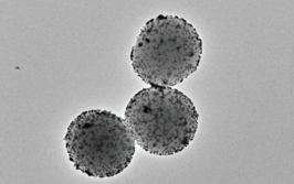
Current methods for fabricating nanoparticles, such as hydrothermal synthesis, laser ablation, or gel synthesis, all involve environmentally unfriendly surfactants, as well as expensive instrumentation. But what if fabrication could be achieved simply with a water bath and hot plate? Inspired by the way water dances on a hot pan – the Leidenfrost phenomenon – and similar chemistry that takes place in underwater volcanos, Mady Elbahri, Professor of Chemical Engineering at Aalto University, Finland, has developed an environmentally friendly means of producing ZnO2 nanoparticles (1). What’s more, Elbahri’s team has also found that the nanoparticles can kill cancer cells. Here, she tells us more about Leidenfrost nanochemistry.
What inspired this work?
Let’s start with the Leidenfrost phenomenon. When cooking in the kitchen, you may have noticed that when a water droplet touches the surface of a very hot pan, instead of evaporating, it moves and dances. I observed this phenomenon – the Leidenfrost phenomenon – in my kitchen a few years ago, and after contemplating the mechanisms behind it, I thought that it could potentially be useful for nanosynthesis. After some initial research, I introduced the novel concept of “Leidenfrost nanochemistry,” which means synthesis of nanoparticles using the Leidenfrost effect. To scale up the process, we sought to recreate the way underwater volcanos form minerals through Leidenfrost chemistry using a hot water bath.
How does Leidenfrost nanochemistry work?
In the proximity of volcano gates, deep in the ocean, the dynamic chemistry taking place is unique in terms of self-regulation and openness to flow conservation, which enables simultaneous chemical synthesis and self-organization of minerals. Similarly, we were able to synthesize nanoparticles at the bottom of a hot bath in an overheated zone at a vapor-liquid interface. The particles then erupt towards the colder region of the liquid-air interface – making them increase in size. This physical separation allows us to tailor the size of the particles with an optimum monodispersity. Monodisperse nanoparticles show uniform properties and induce similar responses in the cells they interact with, which is important in terms of using them for therapeutic purposes.
How do ZnO2 nanoparticles kill cancer cells?
I became interested in ZnO2 nanoparticles after reading the work of Otto Heinrich Warburg, who won the Nobel Prize in 1931 for showing that cancer can be caused by lack of oxygen in cellular respiration. I theorized that peroxide nanoparticles, as a rich source of oxygen, would be able to kill cancer cells by delivering oxygen to cancer cells and inducing oxidative stress. The theory was tested in a series of experiments, which went well. Never before has the impact of ZnO2 nanoparticles on the survival of cancer cells, as well as normal healthy cells, been studied. And the result? ZnO2 nanoparticles have adverse effects on human cells, cancer suspension cells, and adherent tumour cells, depending strongly on the size of the particles and the cell physiology.
What comes next?
We plan to conduct further research to discover what size and dose of the nanoparticles works best for potentially combating cancer. We also hope to learn more about the mechanism involved in the cytotoxic effect of ZnO2 particles on various cell types. We are also looking for investment so that we can further expand the range of applications for Leidenfrost nanochemistry.
- M Elbahri, et al., “Underwater Leidenfrost nanochemistry for creation of size-tailored zinc peroxide cancer nanotherapeutics”, Nature, 12, 15319 (2017). PMID: 28497789.

Over the course of my Biomedical Sciences degree it dawned on me that my goal of becoming a scientist didn’t quite mesh with my lack of affinity for lab work. Thinking on my decision to pursue biology rather than English at age 15 – despite an aptitude for the latter – I realized that science writing was a way to combine what I loved with what I was good at.
From there I set out to gather as much freelancing experience as I could, spending 2 years developing scientific content for International Innovation, before completing an MSc in Science Communication. After gaining invaluable experience in supporting the communications efforts of CERN and IN-PART, I joined Texere – where I am focused on producing consistently engaging, cutting-edge and innovative content for our specialist audiences around the world.
