Fiber Fantastic
Sitting Down with Michael Tanner, Research Fellow in the School of Engineering and Physical Sciences at Heriot Watt University, Edinburgh, UK.
As fiber-optic technology develops at an ever-quickening pace, its applications continue to diversify (1). Now, a multidisciplinary team based in both Edinburgh and Bath, UK, present a new adaptation – a fiber-based screening device intended to aid clinicians treating severe lung conditions by providing in-depth biological information about distal regions of lung tissue (2). We sat down with Michael Tanner, one of the paper’s authors, to find out more…
What was the inspiration behind the project?
The work we published recently is one aspect of a larger project, Proteus, that aims to provide better diagnostic capabilities for people who arrive in intensive care with severe lung problems. There are two main aspects to our work. One is imaging in the lung using optical fibers to observe the presence of bacteria or other pathology-causing agents. In parallel, we are also attempting to better understand conditions in the distal lung because, as things like the acidity or oxygenation levels in these tissues change, they can tell us a lot about the tissue’s health. In fact, our goal is to observe changes in response to treatment. That should help clinicians by producing a more immediate feedback loop.
What are the problems with existing lung disease diagnostics?
Existing diagnostic technology often uses two approaches. The first – X-ray imaging – shows shadows on the lung, but these could be due to inflammation, infection, scarring, or many other causes. It is then possible to take a bronchial lavage (a liquid biopsy) during an endoscopic investigation. This involves delivering fluid into the lung and sucking it out so that it can be analyzed.
The approach is by no means perfect; lavage fluid can easily be contaminated by pathogens in the upper airways, leading to false positives. The technique can also be overly sensitive to pathogens that are not a problem. And the analysis can take time, delaying tailored treatment. In the interim, the clinician may choose to treat broadly, rather than take no action at all.
We have been developing optic fiber technologies to augment diagnosis with immediate feedback to the clinician on infection and tissue health. The technology we’re discussing here is designed with the aim of enabling monitoring using oxygenation and acidity as indicators of disease. We hope to eventually combine this with other techniques to observe the presence of infection – technology toward which we’ve already made great progress (3).
How did you develop the technology?
For us, the real challenge was ensuring that, when we miniaturized the optical fibers, we maintained a strong enough sensor at the end of the fiber. We also wanted to ensure that we could have multiple sensors arranged in a way that avoids overlap. You also need to avoid significant degradation of the sensors during use, and generally come up with an architecture that is small enough to reach a significant depth in the lung while still getting quite a complicated platform into the available space.
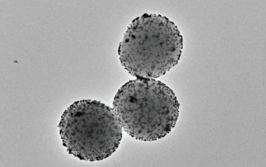
What we’ve managed to do here is establish a way that the chemical sensors can be produced separately from the final probe. They can be produced in a chemical lab – the sensor molecules themselves are fluorescent, and these chemical fluorophores can be attached to very small glass microspheres. On the optical fibers we set up multiple cores as independent, regimented channels. One of the sensors will be attached to each core because we have etched the channels into the end of the fiber.
What have you found out so far about distal lung conditions?
What we’ve demonstrated at the moment is that we are able to take these measurements. We’ve been testing these sensors in various scenarios in ex vivo tissue, but we’ve yet to trial them in live patients. The bench model we’ve been using (whole excised lungs) has confirmed that we are able to see these small changes in the tissue environment – something that wasn’t previously possible.
How will this new technology change the work of those involved in lung diagnostics?
It provides additional information to augment lab-based pathology. Critically, these technologies need investigation alongside current pathology, so that we can tie changes in local oxygenation and acidity in with the progression of disease. For instance, these parameters may improve when a treatment is working, providing reassurance to continue. Miniaturized probes such as ours offer potential for continued monitoring. In the long term, it may be possible for optic fiber-based technologies to reduce the load on lab-based pathologists, or for integration of pathology into the ward via telemetric links to the monitoring instruments.
The key feature of the sensing probe we’ve developed is its small size. You could imagine putting it inside blood vessels, using it in keyhole surgery, or traversing blood vessels to reach organs other than the lungs. We hope that our approach can be applied to other organs – the liver in particular. For now, though, we have focused on the lung because lung diseases are a particularly large burden on health services and a difficult problem to tackle.
The next steps for us are manifold. We’ve not yet fully utilized our platform; although we’ve demonstrated a couple of chemical sensors in this architecture, we’d really like to expand further by measuring not only acidity and oxygen levels, but additional parameters at the same time. Following on from there, it comes back to what we’ve already discussed – demonstrating that monitoring these changes in the tissue can provide clinicians with useful feedback. We’ll need to work in various models to study how different parameters change with tissue health.
What drives you to translate this into the clinic?
It has always been our aim. A major influence on this project has been the pull toward clinical implementation and our desire not to remain stuck in the research lab. We don’t want to be saying, “Oh wouldn’t this be useful, wouldn’t that be useful” forever; we want to actually apply these things.
Our project has taken a very different approach compared to a traditional university research project. As part of Proteus, funded by the EPSRC, we’re working across a number of universities and a number of disciplines. I am by education a physicist, but we also have chemists, biologists, and clinicians working on this. The key to ensuring that we are actually moving forward is the fact that a lot of the work is co-located on-site at the Royal Infirmary of Edinburgh. It allows us to keep focused on making real things happen.
The nice thing about this research work is how well it combines different disciplines. It’s a nice example of the things we are doing to bring different subjects together and to work on something with real-life applications. That’s certainly what’s made us the happiest – getting our work out into the public domain.
- G Keiser et al., “Review of diverse optical fibers used in biomedical research and clinical practice”, J Biomed Opt, 19, 080902 (2014). PMID: 25166470.
- TR Choudhary et al., “High fidelity fibre-based physiological sensing deep in tissue”, Sci Rep, 9, 7713 (2019). PMID: 31118459.
- HE Parker et al., “Fibre-based spectral ratio endomicroscopy for contrast enhancement of bacterial imaging and pulmonary autofluorescence”, Biomed Opt Express, 4, 1856-1869 (2019). PMID: 31086708.

